Putting the science in fiction - Dan Koboldt, Chuck Wendig 2018
Cryopreservation
Star wars and the far future
By Terry Newman
The first thing to remember about cryopreservation is that it’s a very tricky business. The second thing to remember is that frostbite is very painful. Plus, also remember that you have to be very careful about extrapolating from one example—say the freezing of bull semen (no sniggering, please)—to a rather more complicated situation such as people freezing. Some people, when asked what their idea of cryopreservation is, mutter, “That’s all about freezing Walt Disney’s head, isn’t it?” No, it isn’t.
Cryopreservation, as performed by biologists, is actually carried out for two different ends. The first objective is to maintain the structure of biological tissue as close as possible to the functional biological state for subsequent analysis. If you can do this, you might be able to image the biological tissue in as natural state as possible, at a particular time—even capturing cellular events, such as secretion, as they occur and potentially capturing the biochemical constituents of cells and tissues in their appropriate compartments.
The second objective is to permit freezing, and eventual recovery to a fully functional state, of the biological material. This potentially allows for long-term storage, usually at low temperature, with suspended animation. It doesn’t matter if the material, while it is being stored, is in a natural state. A seed, after all, is biological tissue in a form of suspended animation, and it can do remarkably well without going anywhere near a fridge.
These two different approaches to cryopreservation do not go hand in hand. For microscopic and other analytical studies, the cryopreserved biological material should be frozen as quickly as possible, otherwise the fine structure of the cells and tissues will be disrupted by the physical and chemical effects of ice crystal growth. These are not good; in fact, they kill cells. Cryopreserving for future viability is all about controlling the freezing of cellular water to prevent damage and that does not require very fast freezing but controlled freezing.
The truth about liquid nitrogen
Let’s clear up one thing straight away, despite what many novels and Terminator 2: Judgment Day would have you believe: Liquid nitrogen is not a great freezing agent. Yes, it is quite cold (−195.79° C), what we scientists call a bit nippy, but it has quite a small range between its melting and boiling points. This means that if you put something warm into your liquid nitrogen (sitting happily in a vacuum flask), the heat will initially cause the liquid nitrogen to boil. This will form an insulating layer of nitrogen vapor around the object you are trying to cool, which prevents further rapid heat loss.
This is why you can actually very briefly dip your hand in liquid nitrogen (DO NOT TRY THIS AT HOME—OR ANYWHERE!). This is called the Leidenfrost effect. You can see the same thing happen when you put drops of water into a very hot pan.
However, you can get very nasty burns if your clothes are cooled by the liquid nitrogen and then come into contact with your skin. No insulating gas layer is possible then. If you ever have to visit the doctor to get a skin lesion frozen off, the doctor will use quite a high-pressure jet of liquid nitrogen on you. Here the jet effectively strips away the insulating layer of gas to expose the skin to more very cold liquid. Alternatively, you can cool a metal rod in nitrogen and press that against the skin. One way to get around this limiting effect is to make liquid nitrogen slush—a mixture of solid and liquid nitrogen. The heat then removed from the specimen turns the solid into a liquid, rather then the liquid to a gas. This is achieved by putting a small quantity of liquid nitrogen, in an insulated container, into a chamber that can be cooled by a vacuum pump. The resulting liquid nitrogen “Slush Puppie” is a good way of freezing a small amount of biological material and suspensions.
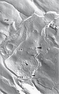
Figure 55.1: An electrically stimulated locust neuromuscular junction frozen milliseconds later using ultra-rapid freezing with a liquid helium-cooled copper block. The sample was then prepared for electron microscopy by freeze-fracture replication, which produces a platinum-shadowed replica of the fractured tissue surface, with white shadows. Membranes can fracture down the center of the bilayer producing an external (E) face and a protoplasmic (P) face. Each of the small particles represents a single membrane protein and is approximately 10nm in size. The large proteins of the pre-synaptic bar (PSB) are thought to be calcium channels. The arrows indicate the position of synaptic vesicles, fused by the freezing process.
EN—External face of the nerve membrane. PM—Protoplasmic face of the muscle membrane. Micrograph prepared by the author.
One of the fastest ways of freezing biological material for ultrastructural analysis is to “slam” it against a liquid helium-cooled ultra-pure copper block. This produces ultrarapid freezing. I used to do this a lot; it’s really cool. (Sorry, I just had to get that pun out of the way.) The electron micrograph shown is of a neuromuscular junction of a locust that was frozen milliseconds after being electrically stimulated. Small membrane-bound vesicles have been captured as they fuse with the nerve membrane. Each of the small particles on the smooth areas is a membrane protein some 14 nanometers in size. A nanometer is 109 meter, or one billionth of a meter. If that doesn’t boggle you, just consider a nanometer is to a tennis ball what the tennis ball is to the Earth. That’s small.
The need for ultrarapid freezing
The objective when freezing for ultrastructural studies is to freeze the tissues so very fast that large ice crystals cannot form to disrupt this delicate nanostructure. The cell water is said to become vitrified—glasslike—and you effectively get a snapshot of the tissue and can capture transient events, like the membrane fusion shown. However, even with the very fast freezing rates that are produced by slamming, you only get this glasslike appearance in a very superficial layer below the contact area (less than 10 micron). This is all because of physics. You simply can’t get the heat out of biological material any faster. And this is the problem with freezing tissue for viability: There is nowhere for the ice crystals to grow where they can’t do damage.
The results of the largely disappointing freezing rates available to us are that you get large ice crystals forming in tissue and they disrupt the structure, bursting membranes and organelles and that will eventually kill cells and tissues—catastrophic for organs! You don’t come back easily from that state when you are put onto defrost. There is another very important effect as well: As water freezes to form pure ice crystals, salts within the cells become concentrated, causing damage thanks to chemical and osmotic effects. If the ice crystals form outside the cells, in the extracellular space, water being drawn out of the cells will cause chemical and osmotic damage within the cells themselves. Slower freezing rates can cause water to freeze outside cells, with the cells themselves dehydrated and damaged by the growing internal salt concentration.
Cryopreservation is therefore a different matter when you are considering freezing for subsequent recovery. That’s all about controlling the freezing rate and the amount and state of the water in the cell.
Cryoprotection of biological tissue
Time for a reminder of some basic biology. The cell is the basic structural and functional unit of any living thing. A tissue is an ensemble of similar cells, usually from the same origin that work together to carry out a specific function. Multiple tissues, working together, form organs. A lot of the work done on cryopreservation for viability has been carried out on isolated cells, like the aforementioned semen and blood components such as red blood cells. Cells are small, and therefore heat can be extracted from them relatively speedily. It is also relatively easy to extract water from, and infiltrate other substances, into a cell. Essentially, this means that isolated cells are ideal for the application of cryoprotectants. These substances are used to protect biological tissue from freezing damage; any discussion about suspended animation is a discussion about the efficacy of cryoprotectants.
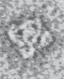
Figure 55.2: An isolated mammalian ryanodine receptor molecule, a type of calcium channel found in the many muscles. The preparation was ultrarapidly frozen in liquid/solid nitrogen slush and viewed under the electron microscope while still frozen. Freezing with cryoprotectants is ideal for this type of investigation. The four clamps at the molecule’s corners and the central rim can be clearly seen. The whole molecule is 270 Å in width. An angstrom is a unit of length equal to 10-10 m. Micrograph prepared by the author.
Cryoprotectants come in two main classes, penetrating and nonpenetrating. Penetrating cryoprotectants are small molecules that dissolve in water and can easily cross cell membranes and affect the melting point of water. These cryoprotectants must not in themselves be toxic, of course, but you need to remember that their actions and toxicity at room temperature will be different than that as they approach freezing. They should also not precipitate or adversely affect the distribution of cell constituents. By entering the cell they will help prevent excessive dehydration of cells; some actually function by forming hydrogen bonds with biological molecules. Sugars such as trehalose and glucose can act as penetrating cryoprotectants.
Nonpenetrating cryoprotectants are large molecules that do not enter the cell—they still need to penetrate a tissue to surround the cells. Not everything is known about how they function, but they seem to have an effect similar to that of the proteins within the cells. Polyethylene glycol and polyvinylpyrrolidone are nonpenetrating cryoprotectants.
Another class of helpful compounds is sometimes referred to as ice blockers, and they can complement, by additions in small amounts, the cryoprotective actions of conventional cryoprotectives.
Existing cryoprotectants
Some cryoprotectants occur naturally. Antarctic fish have evolved cryoprotectant glycoproteins, insects use sugars or plyols, and arctic frogs use glucose, but arctic salamanders can produce glycerol in their livers. Microscopic multicellular water bears (Tardigrada), can survive freezing by replacing most of their internal water with the sugar trehalose.
Cocktails of cryoprotectants have been developed to assist in freezing biological materials, and it’s estimated that we are now on a sixth generation with regard to their success and ability to produce a form of vitrification. Vascular tissue, heart valves, corneas, and cartilage have all been successfully frozen. Progress has also been made on ovary and kidney tissue.
However, when red blood cells are infiltrated with trehalose, maximal survival rates are only in the 90 percent range, with functionality reduced to 60 percent. You can get increased functionality but with a reduction of survival. And remember, 90 percent is in pretty perfect conditions, cells in solutions, not where you need to perfuse a cryoprotectant through a dead body. Whole-body perfusions can be tricky at the best of times—like when the patient is actually alive with a beating heart.
I, for one, would not want to lose approximately ten billion neurons from my brain. Call me old-fashioned, but I don’t really fancy coming back in two hundred years’ time as something akin, at best, to a zombie.